ETH researchers use artificial intelligence to enhance high quality of pictures recorded by a comparatively new biomedical imaging methodology. This paves the way in which in direction of extra correct prognosis and cost-effective units.
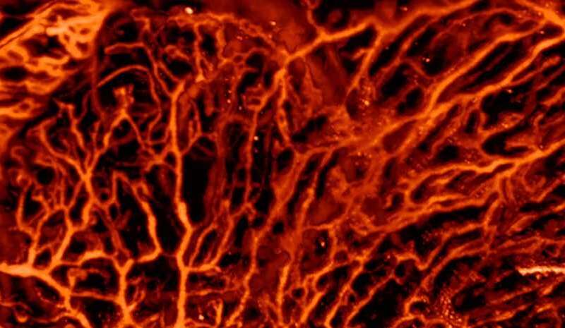
Scientists at ETH Zurich and the College of Zurich have used machine studying strategies to enhance optoacoustic imaging. This comparatively younger medical imaging approach can be utilized for purposes reminiscent of visualizing blood vessels, learning mind exercise, characterizing pores and skin lesions and diagnosing breast most cancers. Nonetheless, high quality of the rendered pictures could be very depending on the quantity and distribution of sensors utilized by the machine: the extra of them, the higher the picture high quality. The brand new method developed by the ETH researchers permits for substantial discount of the variety of sensors with out giving up on the ensuing picture high quality. This makes it doable to cut back the machine price, improve imaging velocity or enhance prognosis.
Optoacoustics (see field) is analogous in some respects to ultrasound imaging. Within the latter, a probe sends ultrasonic waves into the physique, that are mirrored by the tissue. Sensors within the probe detect the returning sound waves and an image of the within of the physique is subsequently generated. In optoacoustic imaging, very brief laser pulses are as a substitute despatched into the tissue, the place they’re absorbed and transformed into ultrasonic waves. Equally to ultrasound imaging, the waves are detected and transformed into pictures.
Correcting for picture distortions
The workforce led by Daniel Razansky, Professor of Biomedical Imaging at ETH Zurich and the College of Zurich, looked for a approach to improve picture high quality of low-cost optoacoustic units that possess solely a small variety of ultrasonic sensors.
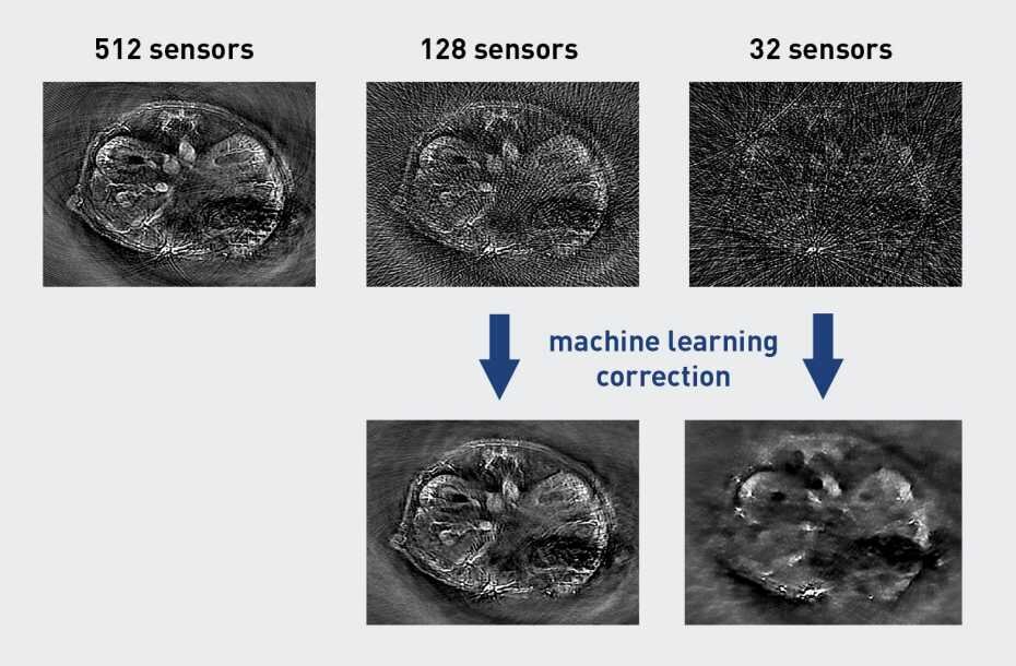
To do that, they began off through the use of a self-developed high-end optoacoustic scanner having 512 sensors, which delivered superior-quality pictures. They’d these footage analyzed by an artificial neural community, which was in a position to be taught the options of the high-quality pictures.
Subsequent, the researchers discarded the vast majority of the sensors, in order that solely 128 or 32 sensors remained, with a detrimental impact on the picture high quality. As a result of lack of knowledge, distortions referred to as streak kind artefacts appeared within the pictures. It turned out, nonetheless, that the beforehand educated neural community was in a position to largely appropriate for these distortions, thus bringing the picture high quality nearer to the measurements obtained with all of the 512 sensors.
In optoacoustics, the picture high quality will increase not solely with the variety of sensors used, but additionally when the data is captured from as many instructions as doable: the bigger the sector wherein the sensors are organized across the object, the higher the standard. The developed machine studying algorithm was additionally profitable in bettering high quality of pictures that had been recorded from only a narrowly circumscribed sector. “That is notably essential for medical purposes, because the laser pulses can’t penetrate the whole human physique, therefore the imaged area is often solely accessible from one course,” in response to Razansky.
Facilitating medical determination making
The scientists stress that their method shouldn’t be restricted to optoacoustic imaging. As a result of the strategy operates on the reconstructed pictures, not the uncooked recorded information, it’s also relevant to different imaging methods. “You’ll be able to principally use the identical methodology to supply high-quality pictures from any form of sparse information,” Razansky says. He explains that physicians are sometimes confronted with the problem of decoding poor high quality pictures from sufferers. “We present that such pictures could be improved with AI strategies, making it simpler to realize extra correct prognosis.”
For Razansky, this analysis work is an efficient instance of what present strategies of artificial intelligence can be utilized for. “Many individuals suppose that AI may exchange human intelligence. That is most likely exaggerated, not less than for the at present obtainable AI know-how,” he says. “It will possibly’t exchange human creativity, but might launch us from some laborious, repetitive duties.”
Of their present analysis, the scientists used an optoacoustic tomography machine custom-made for small animals, and educated the machine studying algorithms with pictures from mice. The following step can be to use the strategy to optoacoustic pictures from human sufferers, Razansky says.
Revealing tissue perform
Not like optoacoustics (often known as photoacoustics), many imaging methods, reminiscent of ultrasound, X-ray or MRI, are primarily appropriate for visualizing anatomical alterations within the physique. To obtain further purposeful data, as an example regarding blood movement or metabolic modifications, the affected person needs to be administered distinction brokers or radioactive tracers earlier than the imaging. In distinction, the optoacoustic methodology can visualize purposeful and molecular data with out introducing distinction brokers. One instance is native modifications in tissue oxygenation—an essential landmark of most cancers that can be utilized for early prognosis. Lipid content material in blood vessels is yet one more potential illness marker, which might support an early detection of cardiovascular ailments.
It ought to be famous, nonetheless, that as a result of the sunshine waves utilized in optoacoustic imaging, not like different waves, don’t totally penetrate the human physique, the strategy is simply appropriate for investigating tissues to a depth of some centimeters beneath the pores and skin.
Novel approach precisely assesses cardiovascular dangers
Neda Davoudi et al. Deep studying optoacoustic tomography with sparse information, Nature Machine Intelligence (2019). DOI: 10.1038/s42256-019-0095-Three
Quotation:
Synthetic intelligence improves biomedical imaging (2019, September 30)
retrieved 30 September 2019
from https://techxplore.com/information/2019-09-artificial-intelligence-biomedical-imaging.html

This doc is topic to copyright. Aside from any truthful dealing for the aim of personal examine or analysis, no
half could also be reproduced with out the written permission. The content material is offered for data functions solely.